
- The microscope is an essential tool for any histology course and even though technology has given us digital microscopes on our computers, it is important to learn the basics of light microscopy and to understand other microscopic instruments.
- Microscopes are specialized optical instruments designed to produce magnified visual or photographic (including digital) images of objects or specimens that are too small to be seen with the naked eye.
- Adding an aberration corrector to a microscope is like fitting it with a pair of glasses, allowing microscopes to see things that they could not see before.
- The magnifying power of a microscope is an expression of the number of times the object being examined appears to be enlarged and is a dimensionless ratio.
- You’ll want to consider things like available outlets and battery availability when deciding which power source on a microscope is right for you.
- The resolution of a microscope is the smallest distance between two objects that results in two images that are distinguishable from each other.
- Microscopes are a ubiquitous tool of science and, in one form or another, are used in laboratories from biomedical to materials engineering.
- A microscope is a piece of scientific equipment that helps us to see things that are invisible to the naked eye, because they are so small.
- Microscopes are effectively just tubes packed with lenses, curved pieces of glass that bend (or refract) light rays passing through them.
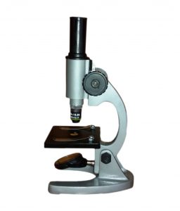
- A microscope is an instrument that can be used to observe small objects, even cells.
- Microscopes are used to resolve detail with sufficient contrast to be made visible.
- The microscope is one of the most important tools used in chemistry and biology.
- Microscopes are designated as either light microscopes or electron microscopes.
- Do remember that focusing a specimen under a microscope is a skill unto itself.
Contents
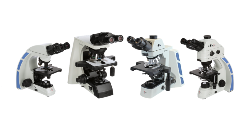
Microscope types:
Brightfield microscopes
A brightfield microscope creates an image by directing light from the illuminator at the specimen; this light is differentially transmitted, absorbed, reflected, or refracted by different structures.
More about Brightfield microscopes…
Comparison microscopes
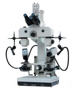
A comparison microscope is composed of two compound microscopes optically joined to provide a real-time split view.
More about Comparison microscopes
Darkfield microscopes
 A darkfield microscope is a brightfield microscope that has a small but significant modification to the condenser.
More about Darkfield microscopes
Digital microscopes
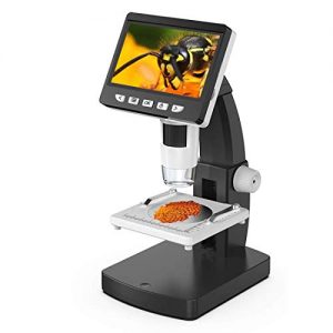 A digital microscope is a microscope equipped with a digital camera allowing observation of a sample via a computer.
More about Digital microscopes
Fluorescence microscopes
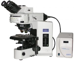 A fluorescence microscope is an optical microscope that uses fluorescence instead of, or in addition to, scattering, reflection, and attenuation or absorption, to study the properties of organic or inorganic substances. “Fluorescence microscope” refers to any microscope that uses fluorescence to generate an image, whether it is a more simple set up like an epifluorescence microscope or a more complicated design such as a confocal microscope, which uses optical sectioning to get better resolution of the fluorescence image.
More about Fluorescence microscopes
Metallurgical microscopes
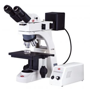 A metallurgical microscope is a compound microscope that may have transmitted and reflected light, or just reflected light.
More about Metallurgical microscopes
Polarizing microscopes
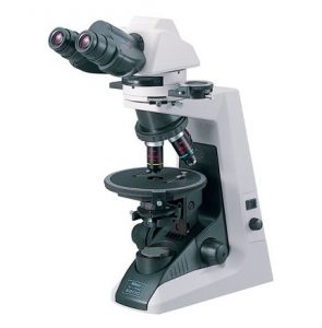 A polarizing microscope is another type of compound microscope.
More about Polarizing microscopes
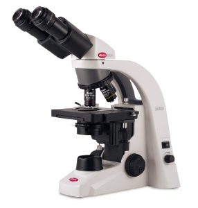
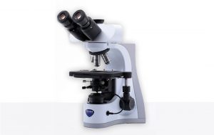
Simple microscopes
 A simple microscope consists of a single lens or several lenses grouped in one unit and are only used to enlarge an object. A simple microscope consists of a single lens that magnifies the object.
History
- In 1625 Stelluti published his Anatomy of the Bee, as Revealed by the Microscope.
- In 1653, Petrus Borellus [1] wrote the first publication on the use of microscope in medicine.
- In 1665, Robert Hooke used an improved compound microscope to observe cells.
- In 1673, with the aid of a crude microscope consisting of a biconcave lens enclosed in two metal plates, Leeuwenhoek introduced the world to the existence of microbial forms of life.
- In 1739 the great Professor Samuel Klingenstierna of the University of Uppsala, Sweden bought a Culpeper-type microscope made by Matthew Loft.
- In 1830, Joseph Jackson Lister created an essentially modern light microscope.
- In 1893 August Köhler developed a key principle of sample illumination, Köhler illumination, which is central to achieving the theoretical limits of resolution for the light microscope.
- In 1932, Ernst Lubcke of Siemens & Halske built and obtained images from a prototype electron microscope, applying the concepts described in Rudenberg’s patent.
- In 1933 a primitive electron microscope was built that imaged a specimen rather than the electron source, and in 1935 Knoll produced a scanned image of a solid surface.
- In 1965, the first commercial scanning electron microscope was developed by Professor Sir Charles Oatley and his postgraduate student Gary Stewart, and marketed by the Cambridge Instrument Company as the “Stereoscan”.
- In 1978 first theoretical ideas have been developed to break this barrier by using a 4Pi microscope as a confocal laser scanning fluorescence microscope where the light is focused ideally from all sides to a common focus which is used to scan the object by ‘point-by-point’ excitation combined with ‘point-by-point’ detection. However, the first experimental demonstration of the 4pi microscope took place in 1994. 4Pi microscopy maximizes the amount of available focusing directions by using two opposing objective lenses or two-photon excitation microscopy using redshifted light and multi-photon excitation.
- In 1986, Binnig, Berger, and Calvin Quate invented the first derivative of the STM—the Atomic Force Microscope.
- In 2005, a microscope capable of detecting a single molecule was described as a teaching tool.